 | 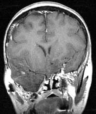 |  |
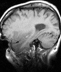
| T1 sagittal pre gad | T1 coronal post gad | T2 axial |
Diagnosis: Neurofibromatosis type I
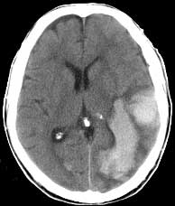
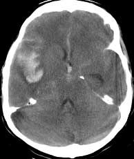
This patient has a history of neurofibromatosis type I and is status post resection of the right eye and some of the sphenoid bone for a plexiform neurofibroma. This is the reason for the herniation of the right temporal lobe into the infratemporal fossa. If you were to look at only the CT images, you might think that there was an aggressive, hemorrhagic neoplasm invading the right middle cranial fossa. However, the MR confirms that there is no abnormal enhancement and that the tissue is actually herniated temporal lobe with hemorrhage from a MCA bifurcation aneurysm. Neurofibromatosis type I is associated with optic nerve gliomas and plexiform neurofibromas. NF2 is associated with meningiomas, astrocytomas, and bilateral acoustic schwannomas. Cutaneous manifestations are common in type I and unusual in type II. NF I represents over 90% of all cases of neurofibromatosis and has autosomal dominant inheritance on chromosome 17. NF I is associated with hypoplasia of the greater wing of the sphenoid, dural ectasia, and cerebral aneurysms among many others. The plexiform neurofibromas of NF I are most commonly associated with the ophthalmic division of the trigeminal nerve. Related Cases