

CT axials pre-contrast


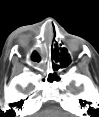
CT axials pre-contrast Findings: Extensive inflammatory changes within the pre-septal soft tissues of the right orbit, with some extension into the medial pre-septal soft tissues of the left orbit. Broad based soft tissue density (a) adjacent to the medial wall of the right orbit displacing the medial rectus laterally. No inflammation of the intraconal fat of the orbits. Right proptosis. Opacification of most of the right ethmoid air cells with a possible bony defect (b) in the medial wall of the right orbit. Apparent air bubble (b) in the medial right orbit is probably the superior extension of the right maxillary sinus. Membrane thickening in the sphenoid and right maxillary sinuses.