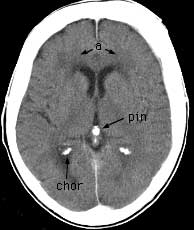


Axial CT post-contrast


Axial CT post-contrast Findings: Generalized thickening of the frontal bones slightly more prominent on the left. Decreased attenuation in the periventricular white matter (a) adjacent to the anterior horns of both lateral ventricles compatible with small vessel ischemic change. M1 segment of the middle cerebral artery (m1). Basilar artery (ba). Superior cerebellar peduncle (scp). Dorsum sellae (dor sel). Sylvian fissure (sylv). Third ventricle (3v). Quadrigeminal cistern (quad). Superior vermis (verm). Calcified pineal gland (pin). Calcified choroid in the atrium of the right lateral ventricle (chor).


