 |  |  |
| T1 post-gad | T2 FSE | FIR |
| | | |
| T1 post-gad | T2 FSE | FIR |
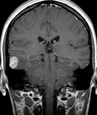
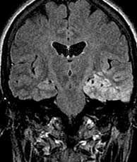
In general, other things to consider in the differential diagnosis of cortically based masses in the temporal lobe include tubers or hamartomas, focal cortical dysplasia, oligodendroglioma, DNET, and ganglioglioma. The appearance of this lesion; however is not good for ganglioglioma since they tend to be more cystic in appearance and often demonstrate enhancement. This patient was otherwise healthy and had no signs of tuberous sclerosis. Related Cases
Heniz ER, Crain BJ, Radtke RA, Burger PC, Friedman AH, Djang WT, Wilkinson WE. MR imaging in patients with temporal lobe seizures: correlation of results with pathologic findings. AJR, Sep 1990; 155(3):p581-6.
Vital A, Marchal C. Loiseau H, Rougier A, Pedespan HM, Rivel J, Vital C. Glial and neuronoglial malformative lesions associated with medically intractable epilepsy. Acta Neuropathol, 1994; 87(2):p196-201.
 |  |  |
| Ganglioglioma | Tuberous sclerosis | DNET |