 |  |  |
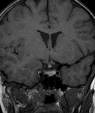
| T1 pre-gad | T1 post-gad | T1 post-gad with fat sat |
| | | |
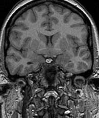
| T1 pre-gad | T1 post-gad | T1 post-gad with fat sat |
The general diagnosis of suprasellar lesions with increased T1 signal pre-gad includes craniopharyngioma, Rathke cleft cyst, hemorrhage, dermoid & lipoma, and ectopic posterior pituitary. Interruption of transport of hormones from the hypothalamus due to such entities as trauma, sarcoidosis or Langerhans cell histiocytosis may also produce a focus of increased T1 signal in the suprasellar region which may mimic these other entities. In this case the differential is limited due to the fact that the lesion is stable over time, mildly enhances and does not contain fat. In addition, the presence of a hypoplastic or absent sella suggests the diagnosis of an ectopic posterior pituitary gland. Related Cases
Kelly WM, Kucharczyk W, Kucharczyk J, et al. Posterior pituitary ectopia: an MR feature of pituitary dwarfism. AJNR, May-Jun 1988; 9(3):p453-60.
Kuroiwa T, Okabe Y, Hasuo K, et al. MR imaging of pituitary dwarfism. AJNR, Jan- Feb 1991; 2(1):p161-4.
el Gammal T, Brooks BS, Hoffman WH. MR imaging of the ectopic bright signal of posterior pituitary regeneration. AJNR Mar-Apr 1989; 10(2):p323-8.
Abrahams JJ, Tefelner E, Boulware SD. Idiopathic growth hormone deficiency: MR findings in 35 patients. AJNR Jan-Feb 1991; 12(1): p155-60.
 |  |  |
| Histiocytosis | Craniopharyngioma | Dermoid |