 | 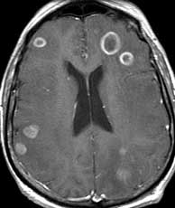 |  |
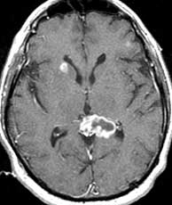
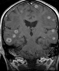
| T1 coronal post gad | T1 axial post gad | T2 axial |
Diagnosis: Metastatic lung carcinoma
40% of all intracranial tumors are metastases and are the most common tumor of the posterior fossa. Lung, breast, GI, renal, and melanoma make up 95% of all brain metastases. 80% of CNS metastases are multiple lesions, usually in the subcortex. Most intracerebral metastases are parenchymal; however, leptomeningeal and dural metastases may also be seen. Metastases are typically round, ring enhancing subcortical lesions with surrounding vasogenic edema which may be identified by decreased signal on T1 and increased signal on T2. Metastases typically have increased T2 signal centrally, however some metastases may have decreased T2 signal. These include mucin secreting tumors like colon carcinoma and other neoplasms with very high ratio of nuclear to cytoplasmic material such as lymphoma. The general differential of multiple ring enhancing lesions includes multiple astrocytomas, mets, toxoplasmosis, cysticercosis, tuberculosis and bacterial abscesses. CNS lymphoma typically presents as a bulky deep white matter or deep gray homogeneously enhancing mass. If the patient is immunocompromised, the findings in CNS lymphoma may be more atypical including central necrosis and ring enhancement. Hydatid disease of brain which is caused by the canine tapeworm echinococcus granulosus is not a differential since it usually presents as a single, well defined cyst with no surrounding edema and little or no ring enhancement. This patient had known metastatic lung carcinoma. This clinical history plus the findings of multiple subcortical ring enhancing lesions with vasogenic edema makes metastatic disease the best choice. Related Cases