 | 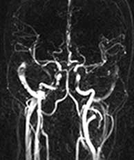 |  |
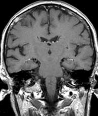
| T1 coronal pre gad | MRA | LCC angiogram AP |
| | | |
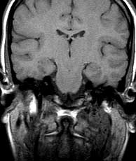
| T1 coronal pre gad | MRA | LCC angiogram AP |
They are intensely enhancing lesions and if they involve the jugular foramen may produce a permeative erosion of the skull base best seen on CT. On non-enhanced MR, they have been described as having a "salt and pepper" appearance due to dark flow voids which are the pepper and small foci of subacute hemorrhage which are the salt. If this pattern is present (not in this case), it is characteristic of paraganglioma.
Schwannoma may present as a carotid space mass. Minimal if any vascularity is present. They have uniform signal without flow voids. Necrosis may be present. If a schwannoma involves a skull base, the erosion of the jugular foramen is more scalloped versus the permeative appearance of a paraganglioma. Likewise, neurofibroma may present as a carotid space mass and is usually indistinguishable from schwannoma. Since the carotid space contains normal lymph nodes, metastases or lymphoma may also present as a carotid space mass.
The location of the mass combined with the clinical history and the vascularity makes glomus vagale the best diagnosis. This patient has a family history of paraganglioma. Related Cases
 |  |
| Breast cancer metastases | Glomus vagale |