|  |  |
| Axials: T1 post gad | PDW | T2 |
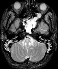
Diagnosis: Hypertrophic olivary degeneration
Hypertrophic olivary degeneration results when there has been interruption of the dentato-rubro-olivary pathways secondary to pontine tegmental or dentate nucleus hemorrhage, infarct, trauma or tumor. It may be contra, ipsi or bilateral depending on the lesion location. It classically presents with palatal myoclonus particularly if bilateral and is characterized by increased signal in the olives which may occur three weeks after the ictus followed by hypertrophy which appears 5 to 15 months later. Histologically there is neuronal degeneration and hypertrophy of astrocytes in the olives. The etiology for this case remains unknown since there was no evidence of infarct, hemorrhage, tumor or history of trauma. Other things to consider which produce increased brainstem signal are multiple sclerosis, central pontine myelinolysis, infarct, trauma, tumor, ascending rhombencephalitis and mitochondrial encephalopathy Related Cases Kithjima M, Korogi Y, Shimomura O: Hypertrophic olivary degeneration MR imaging and Pathologic findings. Radiology August 1994, 192 (2): 539-43.